Lumbar Radiculopathy
Approximately 80% of the population is plagued at one time or another by back pain. Associated leg pain occurs less frequently. Pain can be bothersome and debilitating, limiting daily activities. Leg and back pain can be caused by a variety of reasons, not all of which originate from your spine.
For the purpose of this article, we will focus on lumbar radiculopathy, which refers to pain in the lower extremities in a dermatomal pattern. A dermatome is a specific area in the lower extremity innervated by a specific lumbar nerve. This pain is caused by compression of the roots of the spinal nerves in the lumbar region of the spine. Diagnosing leg and back pain begins with a detailed patient history and examination.
Medical History
Your medical history helps the physician understand the problem. It is important to be specific when answering medical questions related to pain onset but remembering every detail is often not critical. Keeping records of your medical history, including medical problems, medications you are taking and surgeries you have had in the past is helpful.
Regarding your leg and back pain, it may be helpful to keep a journal of your activities, documenting when the pain began, the activities that aggravate your pain and those that relieve your symptoms. It is also important to determine whether your back pain is more bothersome than your leg pain or visa versa. You may be asked if you are experiencing any numbness or weakness in your legs or any difficulty walking. Remember, understanding the cause of your problem is based on the information you provide.
Most people describe radicular pain as a sharp or burning pain that shoots down the leg. This is what some people call sciatica. This pain may or may not begin in the low back. Leg pain caused by compressed nerve roots generally has specific patterns. These patterns of pain depend on the level of the nerve being compressed. After reviewing your history, your physician will perform a physical examination. This will help the physician determine if your symptoms are due to a problem that is caused by spinal nerve root compression. To help you understand the exam performed by your physician lets pause for a quick anatomy lesson.
Anatomy
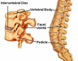
The spine is comprised of 24 vertebrae (bones stacked on top of each other in a “building-block” fashion) that have 4 distinct regions: Cervical, Thoracic, Lumbar, and Sacral. Discs, cushion-like tissues separate most vertebrae and act as the spine’s shock absorbing system. The disc has a tough outer ring of fibers called the Annulus Fibrosus and a soft gel-like center called the Nucleus Pulposus.
There are seven flexible cervical (neck) vertebrae that support the head. There are twelve thoracic (chest) vertebrae, which attach to ribs. The five lumbar vertebrae are large and carry the majority of the body weight. The sacral region helps distribute the body weight to the pelvis and hips.
The spinal cord is housed within the protective spinal column. Spinal nerves come from the spinal cord and travel through a tunnel or foramen. The nerves provide sensory (allowing you to touch and feel) and motor information (allowing the muscles to function) to the entire body.
